The safety of dental X-rays is a common concern that lingers in our minds as we don the lead apron in the dentist’s chair. Amid conflicting information about their necessity and potential risks, it’s time to unravel the truth. In this exploration, we assure you there’s no need to fear the dental detective work involving X-rays.
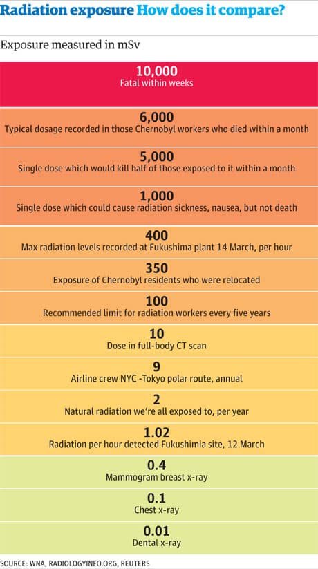
Let’s address the elephant in the room – radiation. Dental x-rays, when conducted with proper precautions, are considered safe. The radiation exposure is minimal, akin to what we encounter in our daily environment. Surprisingly, you’re likely to receive a higher dose of radiation on a flight or basking in the sun than from a dental x-ray. While these X-rays won’t grant you superpowers, they play a crucial role in helping dentists identify potential issues and tailor a personalized treatment plan for your precious smile.
In essence, dental X-rays, with their minimal risk and essential benefits, are a valuable tool in maintaining optimal oral health. Modern technology and protective measures ensure that your exposure is kept to a minimum. If any concerns linger, don’t hesitate to have a candid conversation with your dentist, who can provide further insights and address any worries you may have.

Are Dental X-rays Safe?
Dental x-rays are a common diagnostic tool used by dentists to assess oral health. They provide valuable information about the teeth, gums, and underlying structures that may not be visible during a regular dental examination. However, some people have concerns about the safety of dental x-rays and the potential risks associated with them. In this article, we will explore the safety of dental x-rays and address some common questions and misconceptions.
What are Dental X-rays?
Dental x-rays, or radiographs, are detailed images capturing the structures within the mouth, including teeth, bones, and soft tissues. Utilizing a minimal amount of radiation, these images play a crucial role in detecting and diagnosing various dental conditions such as cavities, gum disease, impacted teeth, and oral infections. They are indispensable for creating precise treatment plans and monitoring oral health over time.
How Do Dental X-rays Work?
Dental x-rays function by emitting controlled radiation through the mouth, capturing images on a digital sensor or photographic film. As the x-ray beam passes through oral structures, it interacts differently with teeth, bones, and soft tissues, creating an image that unveils the internal mouth structures. This process enables dentists to assess the health of teeth and surrounding tissues accurately.
Are Dental X-rays Safe?
Minimal Radiation Exposure: One primary concern is the potential risk of radiation exposure during dental x-rays. However, it’s crucial to note that the levels of radiation used are very low, especially with modern digital systems. The amount falls well within safety limits recommended by regulatory bodies like the American Dental Association (ADA) and the U.S. Food and Drug Administration (FDA).
Comparative Exposure: Radiation exposure from dental x-rays is equivalent to daily background radiation or the amount received during a short airplane flight. The benefits of early detection and prevention of dental issues significantly outweigh the minimal risks associated with radiation exposure.
Precautions Taken: Dentists prioritize patient safety by implementing precautions like lead aprons and thyroid collars to minimize unnecessary radiation exposure during the x-ray process. These measures ensure that the benefits of dental x-rays in maintaining oral health far surpass any potential risks.
Benefits of Dental X-rays
Dental x-rays offer several benefits that contribute to optimal oral health care. Here are some key advantages of dental x-rays:
1. Early Detection of Dental Problems: Dental x-rays can reveal dental issues at their earliest stages, allowing dentists to provide timely treatment and prevent further damage. They can detect cavities, gum disease, cysts, tumors, and other abnormalities that may not be visible during a regular dental examination.
2. Treatment Planning: Dental x-rays help dentists develop comprehensive treatment plans by providing a detailed view of the teeth, roots, and surrounding structures. This helps in accurate diagnosis and allows for effective treatment options.
3. Monitoring Oral Health: Regular dental x-rays are essential for monitoring oral health over time. They help dentists track the progress of dental treatments, identify changes in oral structures, and detect potential problems before they worsen.
4. Precision in Dental Procedures: Dental x-rays assist dentists during complex procedures, such as root canal treatments, tooth extractions, and dental implant placements. They provide valuable information about the position and structure of teeth, ensuring precise and successful outcomes.
Types of Dental X-rays
There are different types of dental x-rays, each serving a specific purpose. The most common types of dental x-rays include:
1. Bitewing X-rays
Bitewing x-rays capture images of the upper and lower teeth in the back of the mouth. They help dentists detect cavities between the teeth, monitor the bone level supporting the teeth, and assess the fit of dental restorations.
2. Periapical X-rays
Periapical x-rays focus on one or two teeth at a time. They provide detailed images of the entire tooth, from the crown to the root, and the surrounding bone. Periapical x-rays are useful for diagnosing dental infections, abscesses, and abnormalities in tooth structure.
3. Panoramic X-rays
Panoramic x-rays capture a wide view of the entire mouth, including the teeth, jaws, sinuses, and temporomandibular joints (TMJ). They are useful for evaluating impacted teeth, jaw disorders, and the overall alignment and development of oral structures.
4. Cone Beam Computed Tomography (CBCT)
CBCT is a three-dimensional imaging technique that provides detailed images of the teeth, bones, and soft tissues. It is commonly used for complex dental procedures, such as orthodontic treatment planning, dental implant placement, and evaluating the temporomandibular joint (TMJ) disorders.
Minimizing Radiation Exposure
While dental x-rays are considered safe, it is still important to minimize radiation exposure whenever possible. Here are some measures taken by dentists to reduce radiation exposure:
1. Use of Digital X-rays: Digital x-ray systems produce significantly lower radiation compared to traditional film-based x-rays. They also offer immediate image results, eliminating the need for chemical processing.
2. Lead Aprons and Thyroid Collars: Dentists provide lead aprons and thyroid collars to patients during x-ray procedures to protect sensitive organs from unnecessary radiation exposure.
3. Proper Technique and Shielding: Dentists follow proper techniques and use shielding devices to ensure the x-ray beam is focused only on the area of interest, minimizing radiation scatter.
4. Individualized Approach: Dentists assess each patient’s specific needs and risks before recommending dental x-rays. They consider factors such as age, oral health history, symptoms, and previous x-ray results to determine the frequency and type of x-rays required.
In conclusion, dental x-rays are a valuable tool in modern dentistry, providing essential information for diagnosing and treating oral health conditions. With proper safety measures in place, the risks associated with dental x-rays are minimal. Dentists prioritize patient safety and follow guidelines to ensure radiation exposure is kept as low as reasonably achievable. Regular dental check-ups, including x-rays when necessary, play a crucial role in maintaining optimal oral health.
Key Takeaways: Are Dental X-rays Safe?
- Dental X-rays are generally safe and necessary for proper dental care.
- The amount of radiation exposure during dental X-rays is minimal and considered safe.
- Pregnant women should inform their dentist to take necessary precautions during X-rays.
- Lead aprons and thyroid collars are used to minimize radiation exposure during X-rays.
- Regular dental check-ups and proper oral hygiene are essential for maintaining dental health.
Frequently Asked Questions
Why are dental x-rays necessary?
Dental x-rays are an essential tool in diagnosing and treating oral health issues. They allow dentists to see areas of the mouth that are not visible to the naked eye, such as between the teeth and below the gum line. X-rays help detect cavities, infections, bone loss, and other dental problems at an early stage, allowing for prompt treatment and prevention of further complications.
While dental x-rays are generally safe, dentists follow the ALARA principle (As Low As Reasonably Achievable) to minimize radiation exposure. They only take x-rays when necessary and use lead aprons and collars to protect the patient’s body from unnecessary radiation.
How often should dental x-rays be taken?
The frequency of dental x-rays depends on various factors, including the patient’s age, oral health history, and risk factors for dental problems. For most adults, bitewing x-rays (which show the upper and lower back teeth) are recommended every 1-2 years, while full-mouth x-rays may be taken every 3-5 years. For children, who are more prone to cavities, x-rays may be taken more frequently.
Your dentist will evaluate your individual needs and determine the appropriate schedule for dental x-rays. It’s important to communicate any changes in your oral health or concerns to your dentist so they can make informed decisions about x-ray frequency.
Are dental x-rays safe during pregnancy?
Dental x-rays are generally safe during pregnancy, especially when appropriate precautions are taken. The American College of Obstetricians and Gynecologists states that dental x-rays, when properly shielded, pose little risk to the developing fetus.
However, it is important to inform your dentist if you are pregnant or suspect you may be pregnant. They will take extra care to minimize radiation exposure by using lead aprons and collars and targeting x-rays only to the necessary areas. If non-emergency x-rays can be postponed until after pregnancy, your dentist may recommend waiting.
Can dental x-rays cause cancer?
The risk of developing cancer from dental x-rays is extremely low. The amount of radiation used in dental x-rays is minimal, and dentists take precautions to protect patients from unnecessary exposure. According to the American Dental Association, the chance of developing cancer from dental x-rays is about one in two million.
It’s important to remember that the benefits of early detection and treatment of dental problems outweigh the minimal risks associated with dental x-rays. If you have concerns about radiation exposure, discuss them with your dentist, who can explain the safety measures taken and address any specific concerns you may have.
Are digital dental x-rays safer than traditional x-rays?
Digital dental x-rays offer several advantages over traditional film x-rays, including reduced radiation exposure. Digital x-rays require significantly less radiation to produce high-quality images compared to traditional x-rays. This makes them a safer option for patients.
In addition to lower radiation exposure, digital x-rays offer faster results, easier storage and retrieval of images, and the ability to enhance and manipulate the images for better diagnosis. They also have a reduced impact on the environment, as they eliminate the need for chemicals used in developing film x-rays.
Are Dental X-rays Safe?
Final Summary: Are Dental X-rays Safe?
Call or Book appointment online
:Ace Dental Care Alpharetta office: 678-562-1555 - Book Now
Ace Dental Care Norcross office: 770-806-1255 - Book Now
Disclaimer
This blog post was generated by artificial intelligence. The content of this post may not be accurate or complete, and should not be relied upon as a substitute for professional advice. If you have any questions about the content of this post, please contact us.
We are constantly working to improve the accuracy and quality of our AI-generated content. However, there may still be errors or inaccuracies. We apologize for any inconvenience this may cause.


